
Osteophytes on Chiropractic X-Rays
An osteophyte is an outgrowth of bone, often called a bone spur. On a spinal X-ray, it may project from a vertebral margin or joint. Its presence alone does not establish the source of pain. Interpret the finding alongside the image, history, and examination, as described in the cervical spondylosis clinical review.
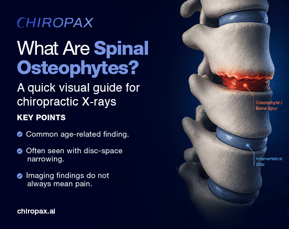
What the radiograph can show
Osteophytes may occur within a broader degenerative pattern. The ABCs of the degenerative spine review describes spondylosis in terms of hypertrophic changes involving vertebral endplates and facet joints. It is a multimodality educational review, not a population-prevalence study. Use it to understand appearances and terminology, rather than to estimate how often a chiropractic patient will have a particular finding.
Record the anatomical level, visible location, and projection on which a finding is seen. Keep that observation separate from a statement about clinical significance. For example, describing a projection along a vertebral margin is different from concluding that it explains a patient's symptoms. When image quality or superimposed structures prevent a confident description, record that limitation instead of forcing a classification.
Read the original studies before quoting percentages
The Brinjikji systematic review pooled 33 studies involving 3,110 asymptomatic individuals. Its inclusion criteria concerned MRI or CT findings. It supports the point that degenerative findings can occur without symptoms, but its estimates are not X-ray osteophyte-prevalence figures. This guide does not transfer its disc-degeneration percentages to bone spurs on plain radiographs.
The Ofiram cervical degenerative index study assessed four-view cervical radiographs from 48 adults who had undergone long fusion for scoliosis. It evaluated a scoring method and observer agreement. Its selected surgical population is not a general chiropractic sample, and the study did not correlate its scores with symptoms. It cannot establish how frequently osteophytes cause neck pain.
The journal record identifies this as a scoring-reliability study. Repeatability within a selected sample does not establish that an imaging sign explains symptoms in another population. Keep the study question attached to the result when discussing the evidence.
Scroll the table horizontally to read all columns.
| Source | Population or method | Appropriate use |
|---|---|---|
| Brinjikji et al., 2015 | Asymptomatic people; MRI/CT systematic review | Context for incidental degeneration, not X-ray osteophyte prevalence |
| Ofiram et al., 2009 | 48 adults after scoliosis fusion; cervical radiographs | Scoring reliability, not symptom causation or general prevalence |
| Kushchayev et al., 2018 | Educational multimodality review | Terminology and appearances, not population estimates |
Keep imaging findings and symptoms distinct
The StatPearls cervical spondylosis review describes osteophytes, disc-space narrowing, and endplate sclerosis among radiographic findings and notes that degenerative imaging changes may correlate poorly with neck pain. That does not make the findings meaningless. It means that an image alone is insufficient to assign a symptom source.
Avoid expanding this statement into a diagnosis or treatment recommendation for an individual reader. A useful report can explain what is visible, where it is located, and what remains uncertain. Decisions about additional assessment belong to the clinician considering the whole presentation. This article does not provide a management protocol or recommend routine imaging for patients without an appropriate clinical indication.
A practical documentation approach
Begin by confirming the study and relevant view. Check whether the region you need to assess is included and whether the image permits the intended observation. Identify the finding before adding software measurements. A nearby measurement line or color does not make a bony projection a symptom-generating lesion.
Separate the record into three ideas: the observation, any measurement actually performed, and the clinician's interpretation. State the method when reporting a measurement. Do not silently turn a qualitative observation into a numerical severity grade, or assign a diagnosis because a software label uses the word significant. If comparisons are made, identify which examinations were compared and whether the views and measurements are comparable.
When explaining the image to a patient, use the same separation. Describe the visible change in plain language, discuss it in the context of the assessment, and be clear about what the image cannot establish by itself. Avoid claims that the size of one spur necessarily predicts the amount of pain or the likely response to care.
Where measurement software fits
ChiroPax's platform overview documents image review, supported AI analysis, manual tools, and reporting. Those functions can organize the technical record. They do not replace interpretation of the underlying image or establish the clinical significance of an individual finding. The clinician reviews the radiograph alongside the assessment.
For a separate angle-measurement topic, see the Cervical Lordosis Measurement Guide. For workflow decisions, use the manual-versus-AI annotation checklist. Ask the ChiroPax team about a specific product output rather than inferring it from a general imaging article.
Frequently Asked Questions
What is an osteophyte?
It is an outgrowth of bone, often described as a bone spur. The degenerative-spine review discusses osteophytes within spondylotic change. Degenerative-spine review
Does an osteophyte on an X-ray prove the cause of pain?
No. The image finding needs clinical context; cervical degenerative findings may correlate poorly with neck pain. Clinical review
Are the Brinjikji percentages X-ray bone-spur statistics?
No. That review's inclusion criteria concerned MRI/CT degeneration findings in asymptomatic people, not plain-film osteophyte prevalence. Original review
Did the Ofiram study establish a link between its scores and symptoms?
No. Its authors explicitly identified the absence of symptom correlation as a limitation. Original study
What should a documentation workflow keep separate?
Keep the visible observation, any measurement actually performed, and the clinician's interpretation distinct. A software label is not a substitute for clinical assessment.
Explore further
Checked on
